In CellProfiler, this is done by thresholding the intensity level in each image. Segmentation means identifying the nuclei in each image.Since we don’t need the colour information, we convert colour images to grayscale type. CellProfiler is designed to work primarily with grayscale images.Metadata is needed to tell CellProfiler what a temporal sequence of images is and what the order of images is in the sequence.Figure 2: Overview of the CellProfiler pipeline using Galaxy tools.ĭetails: More details about the pipeline steps Extract features from the segmented nucleiĪ pipeline is built by chaining together Galaxy tools representing CellProfiler modules and must start with the Starting modules Tool: toolshed.g2.bx.psu.edu/repos/bgruening/cp_common/cp_common/3.1.9+galaxy1 tool and end with the CellProfiler Tool: toolshed.g2.bx.psu.edu/repos/bgruening/cp_cellprofiler/cp_cellprofiler/3.1.9+galaxy0 tool.In this section, we will build a CellProfiler pipeline from scratch in Galaxy. Rename galaxy-pencil the file to drosophila_embryo.zip.Open the Galaxy Upload Manager ( galaxy-upload on the top-right of the tool panel) It is recommended to build a CellProfiler pipeline using the Galaxy interface if the pipeline is to be run by Galaxy. Metadata extraction from file names is limited to a set of fixed patterns.

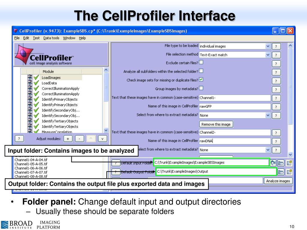
Input and output file locations are set by Galaxy and can’t be set by the user.
#CELLPROFILER SAVE MOVIE NOT WORKING MANUAL#


Combining fluorescent markers with time-lapse imaging is a common approach to collect data on dynamic cellular processes such as cell division (e.g. Most biological processes are dynamic and observing them over time can provide valuable insights.


 0 kommentar(er)
